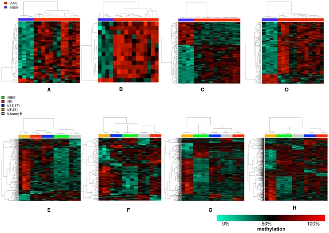
Figure 2. Hierarchical clustering of AML versus NBM in 4 genomic features.
First row represents cluster analysis of all AMLs versus all NBMs and the second row represents cluster analysis of AML subtypes in promoters (A, E), gene bodies (B, F), CGIs (C, G) and CGI shores (D, H). In each figure, each column represents AML patient/NBM and each row represents a single DMR. AML patients were clustered more tightly in CGIs (first row). t(8;21) AML subtype was clustered separately from the other AML subtypes (second row).

