Figure 1.
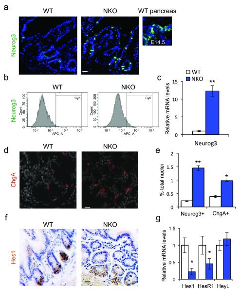
Foxo1 ablation in enteroendocrine progenitors expands the pool of Neurog3+ cells. (a) Neurog3 immunohistochemistry (green) in small intestines of adult WT or NKO mice or in pancreas from developmental day E14.5. (b) Flow cytometry profiles of gut epithelial cell preparations isolated from WT or NKO mice. (c) qPCR analysis of Neurog3 expression in isolated epithelial cells from small and large intestines of NKO (blue bars) or control (white bars) mice. (d) ChgA immunostaining (red) in adult large intestines. (e) Hes1 immunoreactivity (brown) in adult large intestines. (f) Quantification of gut cells reactive with antibodies to Neurog3 or ChgA, a pan-endocrine marker, by flow cytometry analysis. (g) qPCR analysis of Hes1, HesR1, and HeyL expression in isolated epithelial cells from small and large adult intestines. Scale bars: 40 μm (a, f), 30 μm (d), (n = 3 for histology n=4 for flow cytometry, and n = 8 for qPCR). * = P < 0.05, ** = P < 0.01. Error bars indicate SEM.

