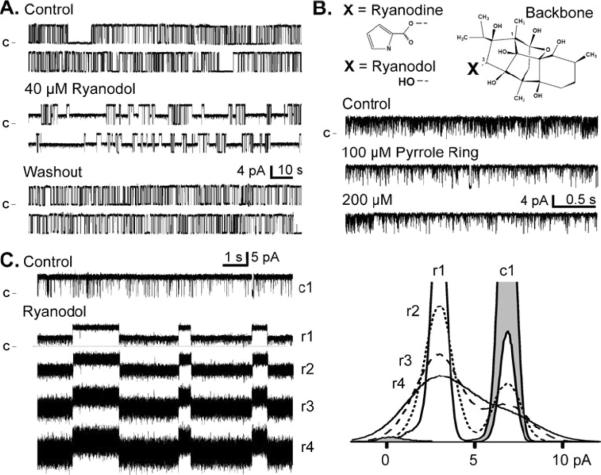
Fig. 1.
Ryanodol modifies single RyR2 function, Openings are upward deflections from the marked closed current level. a Ryanodol action is reversible. All data here are from the same single RyR2 channel recorded at 20 mV. Sample recordings in the top panel are in the absence of ryanodol (control). Sample recordings in the central panel are after 40 μM ryanodol was added to the cytosolic chamber. The bottom panel shows sample recordings after the cytosolic chamber solution was exchanged with a ryanodol-frce solution. The washout period was 5 min long during which 12 volume exchanges were completed. b The ryanoid backbone structure and the 3-C substitutions (X) that produce ryanodine (pyrrole ring) or ryanodol (hydroxyl) are shown (top). Sample single RyR2 channel recordings (bottom) are shown before (control) and after cytosolic addition of the pyrrole ring. c Sample single RyR2 channel recordings without (control) and with 40 μM ryanodol present are shown (left). Here, sample rate was 10 kHz, and trace numbering (c1, r1, r2, r3, and r4) indicates filtering level in kHz. At right are the corresponding all-points histograms (superimposed). The control histogram (c1) is shaded and ryanodol histograms (r1, r2, r3, and r4) are not

