Figure 3.
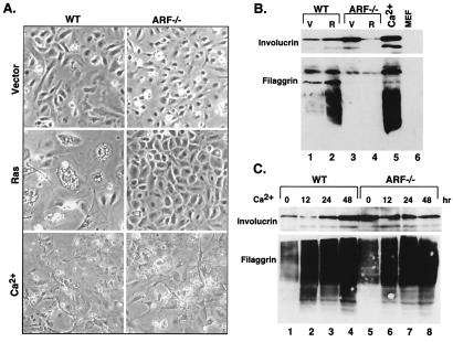
p19ARF is not required for terminal differentiation. (A) Morphological appearance of the cell populations described in Fig. 2. Photographs were taken 48 hr after selection for virus-infected cells, or 48 hr after calcium treatment. (B) Expression of differentiation-associated markers, as shown by using immunoblotting with antibodies directed against involucrin and filaggrin. Extracts from MEFs were used for comparison. V, vector; R, H-rasV12. (C) Expression of differentiation-associated markers in wild-type (WT) and ARF−/− keratinocytes treated with exogenous calcium. Cell extracts were prepared at the indicated times posttreatment, and analyzed by immunoblotting using antibodies directed against involucrin or filaggrin.

