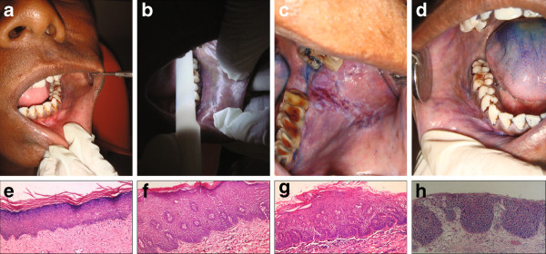
Figure 1.
Study protocol (A-D) and histopathological classification method (E-H). The study protocol included first (A) a regular visual inspection using standard operating procedures and regular light followed by examination using chemiluminescence (B). This was followed by application of toluidine blue. The results of toluidine blue retention were seen as dark royal blue coloration (C) or faintly stained blue coloration (D). Photomicrographs demonstrating the histopathological grading system which classified a lesion as 'no dysplasia' (E) if there was no cell atypia and no changes in architecture; 'mild dysplasia' (F) is there was keratosis, mild cellular atypia and architectural changes in the lower third of epithelium; 'moderate dysplasia' (G) if the architectural changes extended to the middle third of the epithelium; and 'severe dysplasia' (H) if there was a marked cellular atypia associated with architectural changes extending through the entire thickness of epithelium. All photomicrographs show hematoxilin and eosin staining and are depicted at 10× magnification.

