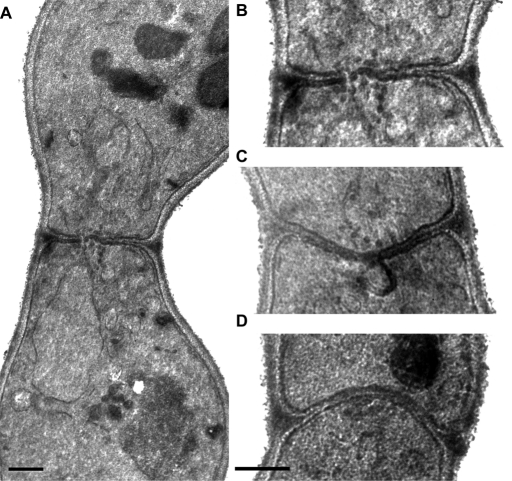
FIGURE 5:
EM of cdc42-138 and fus2 zygotes. cdc42-138 bilateral zygotes (DDY1354 × DDY1355) were examined by electron microscopy. (A) A zygote, showing the unfused nuclei and septum, containing cell wall material. Image is representative of 91 zygotes. (B) The waist regions of the same zygotes, showing vesicles clustered at the zone of cell fusion. Images are representative of 32 zygotes in which vesicles were observed. The vesicles were clustered in 91% of this class. (C) Dark plaques and a membrane invagination at the cell fusion zone. These phenotypes were observed in 21 and 10% of all zygotes examined, respectively. (D) A fus2 zygote, for comparison. Scale bar, 0.5 μm.

