FIGURE 2:
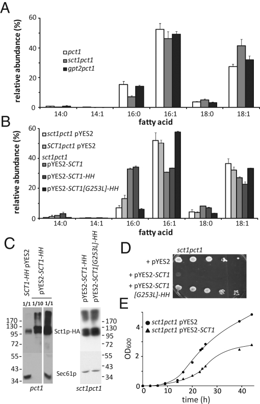
The expression level of catalytically active Sct1p determines the cellular content of C16:0 and affects cell growth. (A) Deletion of the SCT1 gene decreases the cellular C16:0 content. Cells from pct1, sct1pct1, and gpt2pct1 strains cultured in SL to mid-log phase were analyzed for fatty acid content by gas chromatography. (B) Overexpression of SCT1 results in a fourfold increase of C16:0 content dependent on the catalytic activity of Sct1p. Cells from sct1pct1 pYES2 (empty vector control), SCT1pct1 pYES2 (chromosomal expression), sct1pct1 pYES2-SCT1 (overexpression) and sct1pct1 pYES2-SCT1-HH (overexpression of tagged version), and sct1pct1 pYES2-SCT1[G253L]-HH (overexpression of a catalytically dead mutant) strains were cultured to mid-log phase in SGR. In A and B the relative abundance (mol%) of the six major fatty acids is shown, with the error bars representing the SD for sct1pct1 (n = 3), sct1pct1 pYES2 (n = 4), and pct1 pYES2 (n = 5) and the variation for the other strains (n = 2). (C) Western blots comparing the levels of HA-tagged Sct1p chromosomally expressed and episomally expressed from the GAL1 promotor in the pct1 background (left) and of overexpressed catalytically active and inactive Sct1p-HA in sct1pct1 cells (right). Dilution factors of the protein extracts are indicated (left); Sec61p served as loading control. (D) Serial dilutions of the indicated strains precultured in synthetic glucose medium were spotted on SGR agar plates and incubated at 30°C for 3 d. (E) Growth of sct1pct1 pYES2 and sct1pct1 pYES2-SCT1 in liquid SGR medium supplemented with 0.05% glucose. Data averaged from four independent experiments.

