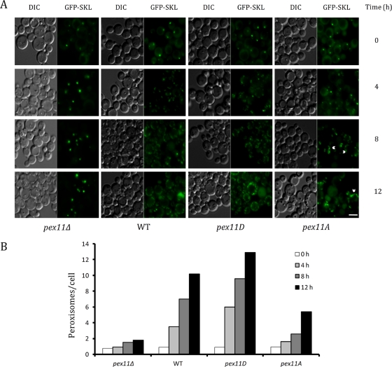
FIGURE 6:
Morphometric analyses of peroxisomes in wild-type and mutant strains. (A) Fluorescence microscopy was performed for pex11Δ, wild-type (WT), pex11D, or pex11A cells expressing GFP-SKL from the GAP promoter. After growth in YPD, cells were transferred to oleate medium and incubated for 12 h. Samples were taken at 0, 4, 8, and 12 h and analyzed by fluorescence microscopy. Arrowheads represent JEPs found in pex11A cells. Scale bar, 4 μm. (B) Graphical representation of the average number of peroxisomes per cell for pex11Δ, wild-type, pex11D, and pex11A cells. Peroxisome counts were performed on 50 randomly recorded cells per time point per strain. Tubulated peroxisomes are counted as single peroxisomes.

