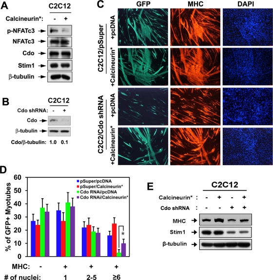
FIGURE 2:
Forced expression of calcineurin restores differentiation of Cdo-depleted C2C12 cells. (A) Lysates of C2C12 cells transfected with a constitutively active form of Calcineurin (Calcineurin*) or control (−) expression vector were subjected to immunoblotting. (B) Lysates of C2C12 cells stably transfected with pSuper or Cdo shRNA were subjected to immunoblotting with antibodies to Cdo and β-tubulin. Cdo and the loading control β-tubulin signals were quantified by densitometry. (C) C2C12/pSuper and C2C12/Cdo shRNA cells were transiently transfected with pcDNA or calcineurin* expression vectors plus a GFP expression vector to mark transfectants. Cells at DM3 were immunostained for MHC (red) and visualized for GFP expression, followed by DAPI staining (blue). (D) Quantification of myotube formation of three independent experiments shown in C. Cultures were scored as MHC negative or MHC positive, with MHC-positive cells further scored as having a single nucleus, two to five nuclei, or six or more nuclei. Values represent means ± SEM from three independent experiments with triple determinations (n = 3). *p < 0.005. (E) Lysates of C2C12/pSuper and C2C12/Cdo shRNA transfected with pcDNA or calcineurin* expression vectors at DM2 were subjected to immunoblotting with antibodies to MHC, Stim1, and β-tubulin.

