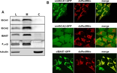
FIGURE 1:
ISCA1, ISCA2, and IBA57 are localized to mitochondria. (A) HeLa cells were treated with digitonin and centrifuged at 15,000 × g to separate the cell lysate (L) into a membrane fraction containing mitochondria (M) and a cytosolic fraction (C). Localization of ISCA1, ISCA2, and IBA57 was analyzed by immunoblotting using antibodies raised against the respective proteins. Antibodies recognizing the α and β subunits of F1-ATP synthase (F1α/β) and tubulin served to estimate the efficiency of separating mitochondrial and cytosolic proteins, respectively. (B) HeLa cells were cotransfected twice with smISCA1-GFP, smISCA2-GFP, or cIBA57-GFP, together with mitochondria-targeted Discosoma sp. red protein (dsRedMito). Images of living cells were acquired by confocal microscopy.

