FIGURE 3:
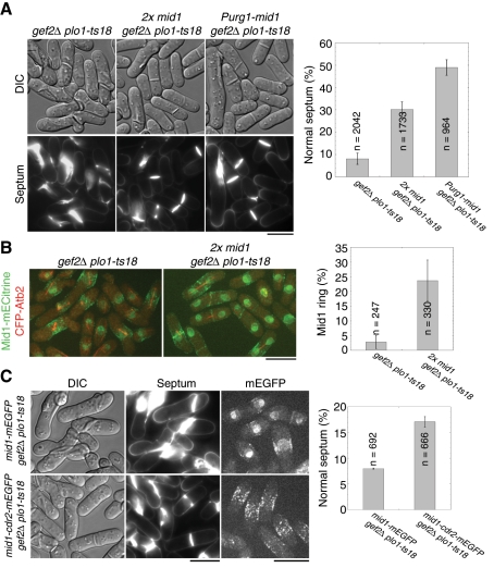
Overexpression of Mid1 rescues gef2∆ plo1-ts18 defects in division-site positioning. (A) Overexpression of Mid1 under different promoters partially rescues septum-positioning defect in gef2∆ plo1-ts18 cells. gef2∆ plo1-ts18 (JW3078), 2x mid1 gef2∆ plo1-ts18 (an extra copy Pmid1-mid1 integrated at the leu1 locus in addition to the native mid1; JW3242), and gef2∆ plo1-ts18 Purg1-mid1 (integrated at the mid1 locus and induced in media with uracil; JW3237) were grown in YE5S medium at 25°C for 24 h. Top, DIC; bottom, calcofluor staining. Septum positioning in calcofluor-stained cells was quantified (right). Mean ± SD from three independent experiments. (B) Two copies of Mid1 partially rescue Mid1 ring formation in gef2∆ plo1-ts18 cells expressing Mid1-mECitrine and CFP-Atb2. Strains JW3281 and JW3383 grown at 25°C. The percentage of mitotic cells (with a spindle) with a Mid1 ring was quantified from two experiments. (C) Mid1-Cdr2 fusion protein partially rescues septum-positioning defect in gef2∆ plo1-ts18 cells. Left, DIC; middle, calcofluor staining; right, Mid1-mEGFP (top, JW3560) or Mid1-Cdr2-mEGFP (bottom, JW3426). Septum positioning in calcofluor-stained cells was quantified from two experiments. Bars, 10 μm.

