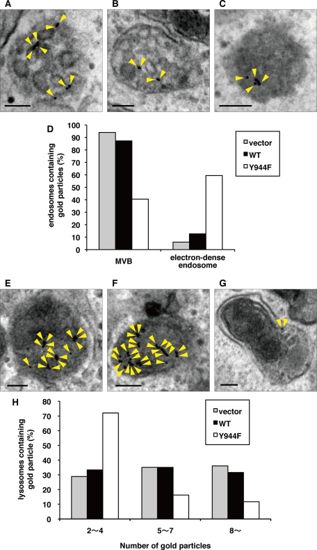
FIGURE 8:
LRRK1(Y944F) fails to form proper intraluminal vesicles of MVBs. (A–C, E–G) We generated HeLa S3 cells stably expressing GFP (A, E), WT GFP-LRRK1 (B, F), and GFP-LRRK1(Y944F) (C, G) according to the manufacturer's protocol. After serum starvation, cells were incubated with anti-EGFR antibodies (LA-22) for 2 h at 37°C, followed by goat anti–mouse IgG antibodies conjugated to 10-nm gold for 1h at 37°C. After washing to remove antibodies from the medium, cells were stimulated with EGF for 10 min (A–C) and 30 min (E–G) and then fixed. The localization of EGFR was examined by conventional electron microscopy. Arrowheads indicate EGFR labeled with immunogold particles. Scale bars, 100 nm. (D) Histogram indicates the percentage of compartments (MVBs or electron-dense endosomes) containing gold particles compared with total endosomes containing gold particles after 10 min of EGF uptake. About 100 organelles that contained gold particles were counted per samples. At this time point, 64% (vector), 60% (LRRK1 WT), and 63% (LRRK1(Y944F)) of organelles were localized in the endosomal compartments. (H) Histogram indicates the percentage of lysosomes containing the indicated number of gold particles after 30 min of EGF uptake. Electron micrographs of ∼100 lysosomes were obtained per sample, and the total number of gold particles was counted for each lysosome. Lysosomes containing more than two gold particles were counted.

