
Fig. 1.
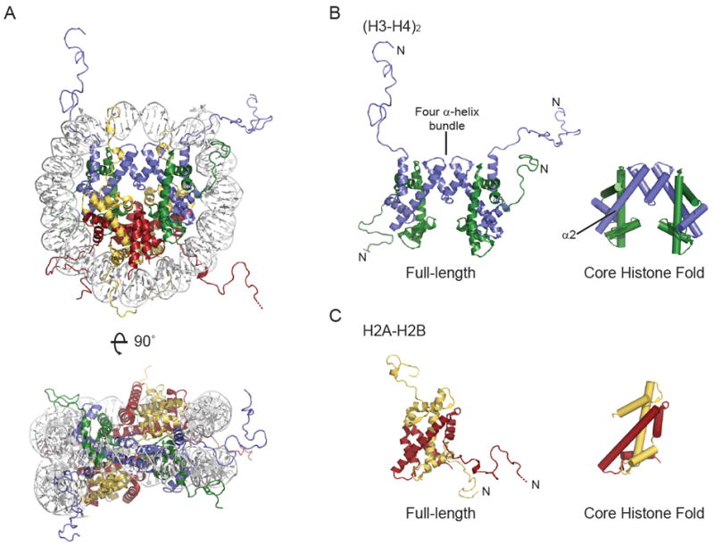
Structure of the major-type nucleosome (A), (H3-H4)2 tetramer (B) and H2A-H2B dimer (C). Cartoons were made using PDB 1KX5. DNA is white, H3 is blue, H4 is green, H2A is yellow and H2B is red. Missing residues at the N-terminus of H2B are shown by a dotted line. In B and C, an N indicates the proteins N-terminus, and the representation on the right shows only the core histone fold. The four α-helix bundle that mediates H3-H4 tetramer formation and the second α-helix of H3 are indicated.