
Figure 2.
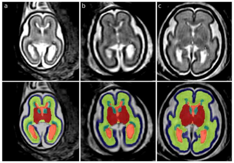
T2w MRI axial sections in native orientation of fetal brains at (a) 21.00, (b) 23.71 and (c) 27.86 gestational weeks (GW) are shown in the top panel. In the occipital lobe, the germinal matrix (GMAT, 1) rapidly thins in the posterior direction and the outer subventricular zone (OSVZ, 2) becomes more cell dense. The GMAT, OSVZ, and subplate (3) are therefore labeled in the top panel to show that the hypointense part around the occipital horn is OSVZ rather than GMAT. In the bottom panel, automated segmentations are overlaid on axial sections: blue, cortical plate (CP); yellow, subplate, intermediate zone and outer subventricular zone (SP+IZ); turquoise, ventricular zone, inner subventricular zone and ganglionic eminence (GMAT); red, deep gray nuclei (DG); orange, lateral and third ventricles (VENT).