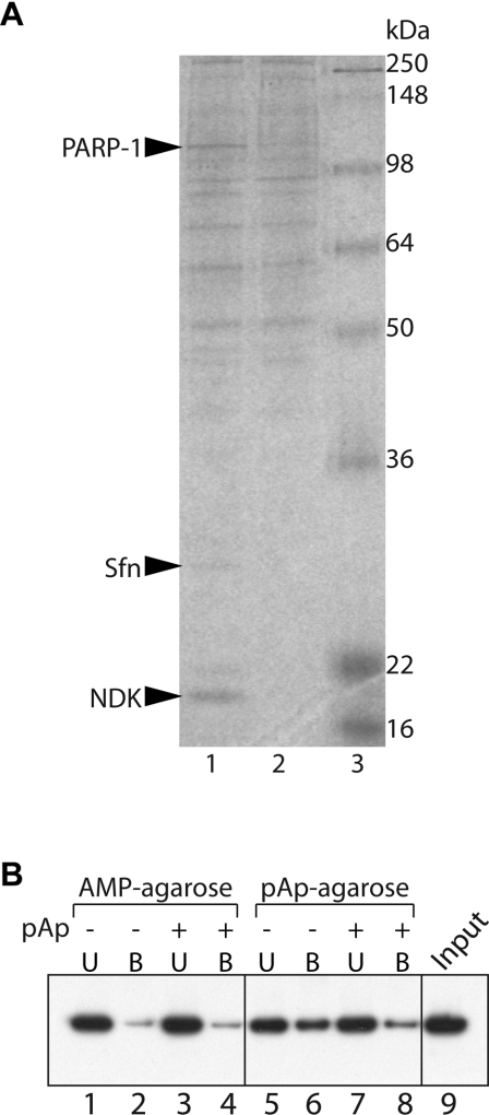
Figure 1. Physical interactions between pAp and PARP.
(A) SDS/PAGE of pAp-affinity chromatography from HeLa cell nuclear extracts. The gel was stained with Colloidal Blue. Lane 1, pAp-binding fraction; lane 2, binding on control agarose beads; lane 3, molecular mass markers. (B) PARP-1-affinity chromatography with AMP-agarose or pAp-agarose. Partially purified PARP-1 (1 μg) was incubated with AMP-agarose (lanes 1–4) or pAp-agarose (lanes 5–8) in the presence (+) or absence (−) of 3 mM pAp. Detection of PARP-1 in the bound (B) or unbound (U) fractions was performed by Western blot analysis. Lane 9, PARP-1 input.

