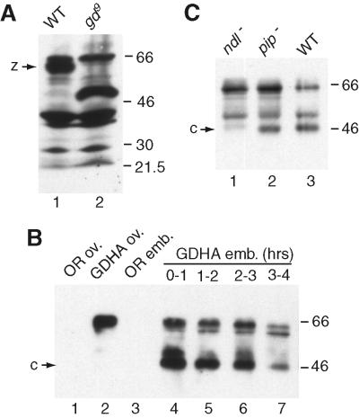
Figure 4.
Cleavage of GD in vivo. (A) Anti-GD Western blot of ovary extracts from wild-type (WT; lane 1) and gd9 (lane 2) mutant females. GD zymogen (z) is absent and replaced by a smaller polypeptide in gd9. (B) Time course of GDHA cleavage. Shown are immunoprecipitations with anti-HA antibody of extracts from ovaries (lanes 1 and 2) or laid eggs (lanes 3–7) at indicated times of embryonic development (lane 3 = 0–4 h), which have been probed on Western blots with anti-GD. The same band pattern is seen when the antibodies are used in reverse order (not shown), indicating that the 46-kDa band (c) represents a C-terminal GD fragment. (C) The 46-kDa form of GDHA (c) is seen in wild-type (WT; lane 3) and pip− (lane 2) but not in ndl− (lane 1) background. Immunoprecipitations of egg extracts and blots were performed as in B.

