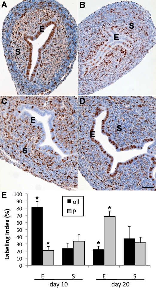
FIG. 3.
Effects of neonatal P4 treatment on uterine cell proliferation as measured by immunohistochemical staining of MKI67. At PND 10, extensive proliferation was seen in both epithelium (E) and stroma (S) of control uteri (A). In contrast, uterine epithelial proliferation in 10-day-old mice treated with P4 during PNDs 3–9 was dramatically reduced (B), although stromal (S) proliferation remained robust. By PND 20 in control uteri, both epithelial and stromal proliferation (C) was reduced compared to PND 10. In contrast, 20-day-old uterine epithelium in mice that received P4 during PNDs 3–9 showed robust epithelial proliferation (D) exceeding that of controls. Bars = 100 μm. Quantitation of uterine epithelial and stromal labeling index in the groups mentioned above indicates that epithelial proliferation in mice treated during PNDs 3–9 with P4 is sharply reduced compared to controls at PND 10 but significantly exceeds controls by PND 20 (E). Stromal labeling index was not significantly different than control in P4 (P)-treated mice at PNDs 10 or PND 20, despite a trend toward an increase in the former. *P < 0.05 vs. control. Data are shown as mean ± SEM.

