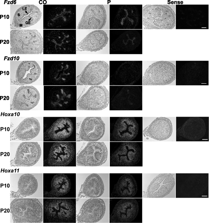
FIG. 6.
Spatial localization of Fzd and Hox gene expression was examined by in situ hybridization analysis in uteri from control mice (CO) and mice that received P4 (P) injections daily during PNDs 3–9 (n = 5 per treatment per day). In each panel portion, representative photomicrographs of in situ hybridization results are shown with light-field and dark-field illumination, respectively. GE, glandular epithelium; LE, luminal epithelium; M, myometrium; S, stroma. Bars = 100 μm.

