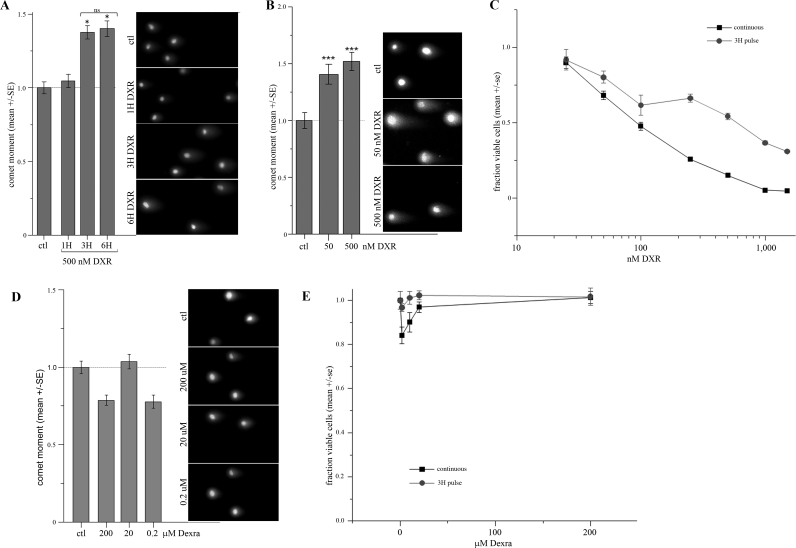
FIG. 1.
Acute DNA damage and cytotoxicity profiles of DXR and Dexra in KK-15 cells. A) KK-15 cells were treated with 500 nM DXR for 1, 3, and 6 h, and the cells processed for the NCA. OM measurements demonstrate ∼45% increase in DNA damage over control at 3- and 6-h treatments (n = 3, *P < 0.01, one-way ANOVA). B) Treatment with 50 or 500 nM DXR for 3 h induced a 40%–50% increase in DNA damage (OM) in the NCA (n = 5, one-way ANOVA, P < 0.005). C) Treating KK-15 cells with a 3-h DXR pulse induced a dose-dependent cytotoxicity response that was less severe than but parallel to continuous exposure (two-way ANOVA, P < 0.001). The fraction viable cells was assessed by mean ATP-based luminescence (CellTiter-Glo assay) normalized to control (n = 3). D) Treatment with 0.2–200 μM Dexra for 3 h did not induce any DNA damage (OM) in KK-15 cells over the controls (DMSO carrier treated) as measured by the NCA (n = 4, one-way ANOVA, P > 0.05). E) Dexra did not cause significant toxicity in KK-15 cells. The fraction viable cells plotted for 3-h pulse or continuous 24-h exposure to Dexra (n = 3). Original magnification ×200 (A, B, D).

