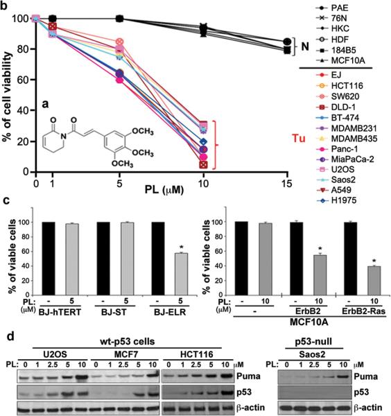
Figure 1. Selective killing effect of PL in cancer cells by a small molecule.
a, Structure of PL. b, PL treatment induces cell death in cancer cells, but does not induce cell death in normal cells. Human normal cells, including aortic endothelial cells (PAE), breast epithelial cells (76N), keratinocytes (HKC), and skin fibroblasts (HDF), as well as two immortalized breast epithelial cell lines (184B5 and MCF10A), were grown in 12 or 24 well plates and treated with PL at 1–15 μM for 24 h. Cytotoxicity was measured by trypan blue exclusion staining (average of three independent experiments). A variety of human cancer cell lines were treated with PL or DMSO (control) for 24 h. PL was HPLC-purified (~99% purity) prior to the treatment. c, Selective cell death by PL in oncogenically transformed human BJ skin fibroblasts (the left panel) and MCF10A cell lines (right panel). A representative graph for cell viability is shown (mean ± SD of three independent experiments; *p<0.0001). d, The effects of PL on p53 and its target PUMA, and pro-survival proteins were measured by western blot analyses in several cancer cells.

