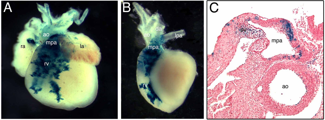
Fig. 5. The fate of Tbx1-expressing cells during late cardiac development.
(A) Frontal view of Tbx1-Cre/R26R embryonic heart at E17.5. (B) Left lateral view of the same heart after removing atria. (C) Transverse sections of the same heart at the pulmonary valve (pv) level. LacZ-positive cells were localized in the anterior portion (outflow tract) of the right ventricle (rv) and the main trunk of the pulmonary artery (mpa) (A, B). LacZ-positive cells were detectable in both endothelial and muscle layers of mpa as well as pv (C). Few blue cells were observed in the wall of aorta (ao in C). ra, right atrium; la, left atrium; da, ductus arteriosus.

