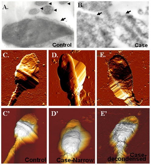
Figure 6. Spermatozoa from c.474A/A patients with abnormal head shape.
(A.–B.) TEM images of sperm isolated from a fertile control (A.) and an infertile mam with c.474A/A (B). The latter shows de-condensed chromatin. Arrows indicate the nucleus; arrow heads indicate the axonemal 9+2 structures (Magnification: ×10,000). (C.–E.) Top-view AFM images confirm abnormal morphology in sperm head. Sperm of a control subject (C.). Sperm of an infertile man with c.474A/A have a narrow head (D.) or a de-condensed nucleus (E.). Three-dimensional images are displayed in the bottom (C′.–E′.).

