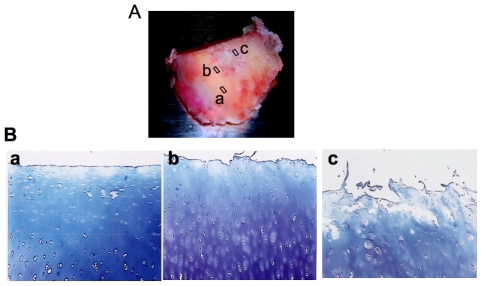
Figure 1. Representative sample of femoral condyle from human OA knee and toluidine blue stained sections.
(A) Femoral condyle from human OA knee joint indicating three sites biopsied for further biochemical and molecular analysis; (a) cartilage sample taken from macroscopically unaffected area and distant from the lesion was termed “normal” (b) cartilage sample isolated from areas immediately close to lesion was termed “Next to lesion”; (c) cartilage specimen excised from lesion was termed “late-stage OA”; Cartilage samples were isolated using a 6 mm-biopsy punch. (B) Representative photomicrographs of cartilage sections showing areas from where samples were recovered for toluidine blue staining; (a) cartilage section from a normal area showing a relatively smooth articular surface; (b) section from areas close to lesion showing dense staining for PGs in mid zone of the cartilage where chondrocyte proliferation and activation occurs; (c) cartilage section from lesions showing degraded articular surface, loss of PG staining and chondrocyte cloning (original magnification ×40).

