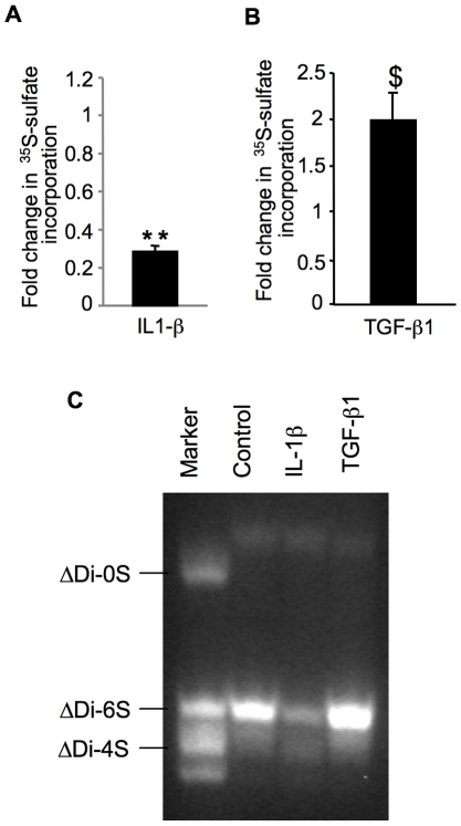
Figure 6. Analysis of the effect of IL-1β and TGF-β1 on GAG synthesis and GAG chain composition.
Cartilage explants from normal regions of human femoral condyle were exposed to IL-1β (A) or TGF-β1 (B) for 24 h and then GAG synthesis was measured by 35S-sulfate incorporation. Measurements were normalized to control (non treated). Data are mean ± SD of 3 experiments per parameter, per joint, per patient. **Significantly (P<0.0018) lower than controls; $significantly (P<0.05) higher than controls. (C) Effect of IL-1β and TGF-β1 on GAG chain composition in human cartilage. Cartilage samples derived from normal regions of human femoral condyle were treated with cytokines for 12 h and analyzed by FACE. Gels are representative images from 3 experiments per parameter, per joint, per patient. Markers are ΔDi0S, nonsulfated chondroitin-unsaturated disaccharide; ΔDi4S, chondroitin-4-sulfated unsaturated disaccharide; ΔDi6S, chondroitin-6-sulfated unsaturated disaccharide.

