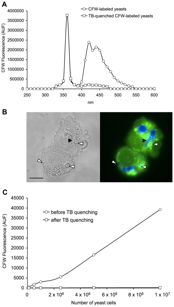
Figure 1. Trypan blue efficiently quenched CFW-labeled yeast fluorescence.
(A) Emission spectrum of CFW-labeled yeasts before and after trypan blue (TB) quenching. 1×10⁷ C. lusitaniae cells were labeled with 5 µg/ml CFW, and 250 µg/ml trypan blue was used for quenching. Excitation wavelength was set up at 360 nm and emission wavelength was set up to cover the entire emission spectra of CFW. (B) Macrophages after 4 hours of infection with C. lusitaniae. Yeast cells were stained with CFW (blue), macrophages were stained with calcein (green). Left panel: phase contrast, right panel: fluorescence. The fungal cells ingested inside the macrophages were protected from trypan blue quenching and were fluorescent, whereas membrane-bound yeasts exposed to trypan blue quenching were not (some examples are shown with white arrowheads). A pseudohyphae of C. lusitaniae can be seen within the macrophage (black arrowhead). The bar represents 20 μm. (C) Fluorometric detection of CFW-labeled C. lusitaniae cells before and after trypan blue quenching. Trypan blue used at 250 μg/ml quenched the CFW fluorescence to background level for up to 1×10⁷ C. lusitaniae cells.

