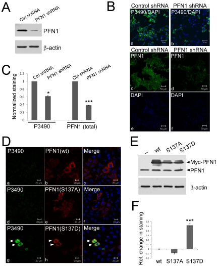
Figure 2. P3490 specifically detects pS137-PFN1 via immunofluorescence staining.
A, HEK293 cells were infected for three days with lentiviral shRNA particles encoding either a scrambled nucleotide sequence (Ctrl shRNA), or a target-specific sequence for human PFN1 (PFN1 shRNA). Efficient PFN1 knockdown was confirmed by Western blot. B, virus-mediated PFN1 knockdown reduced P3490 staining (green, a–b), as visualized by confocal fluorescence microscope. Total PFN1 decrease was also confirmed by immunofluorescence staining (green, c–d). DAPI (blue) was used to counterstain cells either as merged (a–b) or unmerged images (e–f). C, staining of virus-infected cells with P3490 and PFN1 antibodies (as in A and B) was quantified by fluorescence plate reader, and normalized vs. DAPI to control for cell numbers. The decrease of P3490 (∼40%; *, p<0.05, unpaired t-test) and total PFN1(60%; p<0.001, unpaired t-test) staining caused by PFN1 knockdown was calculated relative to cells infected with the control shRNA (arbitrarily set as 1). Error bars represent the standard error of the mean (SEM). Data are mean ± SEM of three independent experiments. D, HEK293 cells were transiently transfected with Myc-tagged PFN1(wt, S137A or S137D), treated with 50 µM hydroxyfasudil for 16 hr, and double stained with an anti-Myc (red) and P3490 (green) antibodies. Only cells expressing the phosphomimetic Myc-PFN1(S137D) stained positive with P3490 (g–i), but not Myc-PFN1(wt) (a–c) or Myc-PFN1(S137A) (d–f). Arrowheads indicate two cells that expressed Myc-PFN1(S137D) and stained positive with P3490. E, Western blot confirmed comparable levels of Myc-PFN1 over-expression (wt, S137A and S137D) in transiently transfected HEK293 cells, as compared to cells transfected with an empty vector (pcDNA3). F, cells transfected (as in E) with pcDNA3 or various PFN1 constructs were immunostained with P3490, and quantified on a fluorescence plate reader with normalization vs. DAPI. The effects of PFN1 over-expression on P3490 staining are represented as relative change compared to cells transfected with pcDNA3. Only PFN1(S137D) increased P3490 staining (***, p<0.001, unpaired t-test). Cells were not treated with hydroxyfasudil. Error bars represent the SEM. Data are mean ± SEM of three independent experiments.

