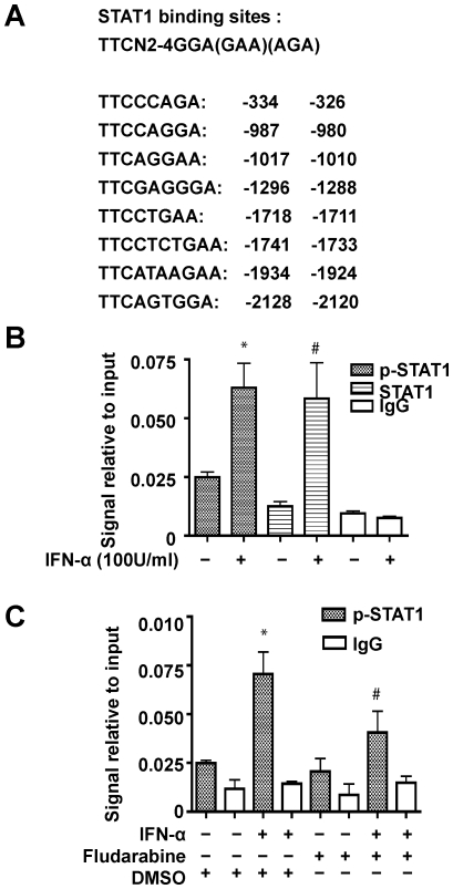
Figure 4. STAT1 binds directly with the GLS1 promoter in IFN-α treated MDM.
(A). The predicted STAT1 binding sites in the human GLS1 promoter, TSS is designated as +1. (B). MDM were treated with 100 U/ml IFN-α for 1 hour, then ChIP assay was performed using digested chromatin, p-STAT1 (Tyr 701) and STAT1 antibodies, or IgG antibody as a negative control. Purified DNA was analyzed by quantitative real-time PCR using specific primers. The amount of immunoprecipitated DNA is represented as signal relative to the total amount of input chromatin. The data are representative of three independent experiments using three different donors. #, p<0.05, *, p<0.01 in comparison with control. (C). MDM were pretreated with 1 µM fludarabine or 1:10,000 DMSO for 1 hour, then treated with or without 100 U/ml IFN-α for another hour. ChIP assay was performed using p-STAT1 antibody as in (B). The data are representative of two independent experiments using two different donors. *, p<0.01 in comparison with control that were treated with DMSO only; #, p<0.05 in comparison to cells treated with IFN-α and DMSO.

