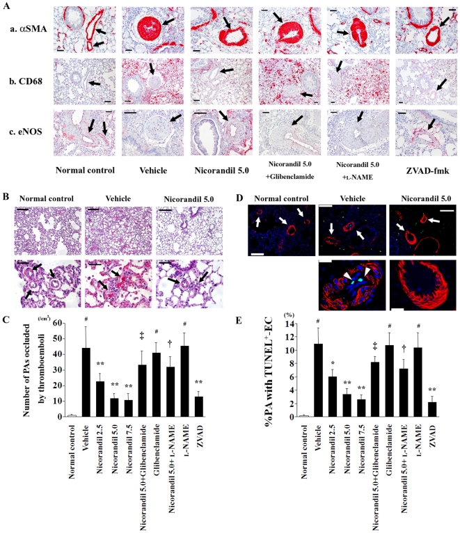
Figure 2. Nicorandil improves the histopathological findings of MCT-injured lungs.
(A) Immunohistochemical staining for αSMA (a), CD68 (b), and eNOS (c) in lung sections are shown. Histopathologically, MCT injury induced the thickened medial wall of PAs (arrows) that was composed of αSMA-positive cells, the recruitment of CD68-positive macrophages into the perivascular areas, and the deficiency of eNOS expression in the endothelium of the pulmonary vasculature (see vehicle). Nicorandil and ZVAD-fmk improved these deleterious histopathological changes in the MCT-injured lungs, whereas glibenclamide and l-NAME blocked the effects of nicorandil. Scale bar, 50 µm. (B) MCT readily induced thromboemboli formation that occluded the small PAs (arrows; see vehicle), which was attenuated by nicorandil. HE staining. Scale bars, 100 µm (top) and 50 µm (bottom). (C) The number of small PAs that were occluded by thromboemboli was counted in the lung sections. (D) TUNEL staining with immunofluorescence staining for αSMA in lung sections. MCT readily induced endothelial cell apoptosis (arrowheads) in a number of PAs (arrows; see vehicle), which was attenuated by nicorandil. TUNEL (green); αSMA (red); and nuclei (blue). Scale bars, 100 µm (top) and 20 µm (bottom). (E) The number of small PAs having TUNEL-positive endothelial cell(s) [EC(s)] was counted in lung sections, and the proportion of PAs with TUNEL-positive EC(s) in a total of equally sized PAs was calculated. # P<0.01 vs. normal control; * P<0.05 and ** P<0.01 vs. vehicle; † P<0.05 and ‡ P<0.01 vs. nicorandil (5.0 mg·kg−1·day−1).

