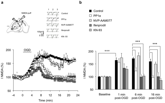
Figure 5. PP1α expression and CaMIIα inhibition normalize NR2B-containing NMDAR-mediated currents after excitotoxicity.
(a) Significant enhancement of NMDAR currents following 4 min OGD in control slices (control, n = 6). Expression of PP1α (n = 5), ifenprodil treatment (n = 8), and KN-93 treatment (n = 6) led to a full recovery of NMDAR currents 6 min after the end of OGD. NVP-AAM077 (n = 5) has no effect on NMDAR currents that remain potentiated throughout recording. Top left, Schematic representation of an organotypic hippocampal slice showing the positioning of the recording patch-clamp electrode and the puffing pipette. Top right, Individual responses from single CA1 pyramidal neurons before (1), during (2) and 10 min after (3) OGD. (b) Time course of OGD mean effect on NMDAR currents. +++p<0.001, ***p<0.001.

