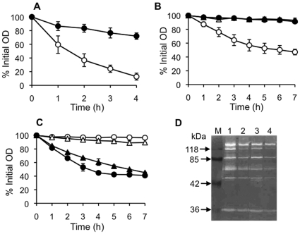
Figure 4. Effect of amicoumacin A on autolysis.
(A) Effect on whole cell autolysis. Cultures grown in the absence (open circles) and the presence (closed circles) of amicoumacin A were washed and suspended in autolysis buffer to an initial OD600 of around 1.0 and autolysis was monitored as decline in OD600. (B) Quantitative assay of murein hydrolase activity against cell wall. An equal amount (90 µg protein) of extracellular proteins were added to cell wall purified from amicoumacin-A-untreated cells and OD600 was monitored as described in Materials and Methods. Symbols: open circles, extracellular proteins from untreated cells; closed circles, extracellular proteins from amicoumacin-A-treated cells; open triangles, 10 mM Tris-HCl (pH 7.5). (C) Susceptibility assay of purified cell wall to murein hydrolase. Ninety µg of extracellular proteins from amicoumacin-A-untreated cells were added to cell wall purified from amicoumacin-A-treated and -untreated cells and OD600 was monitored. Symbols: open circles, cell wall from untreated cells with 10 mM Tris-HCl (pH 7.5); closed circles, cell wall from untreated cells with extracellular proteins; open triangles, cell wall from amicoumacin-A-treated cells with 10 mM Tris-HCl (pH 7.5); closed triangles, cell wall from amicoumacin-A-treated cells with extracellular proteins. (D) Zymographic analysis of murein hydrolase activity from amicoumacin-A-treated and -untreated cells against purified S. aureus COL cell wall. Autolytic extracts were prepared and assayed by electrophoresis on an SDS-polyacrylamide gel (10%) containing 1 mg/ml purified cell wall as described in Materials and Methods. Lanes: M, prestained molecular weight markers; 1, cell-wall-associated proteins from untreated cells; 2, cell-wall-associated proteins from amicoumacin-A-treated cells; 3, extracellular proteins from untreated cells; 4, extracellular proteins from amicoumacin-A-treated cells.

