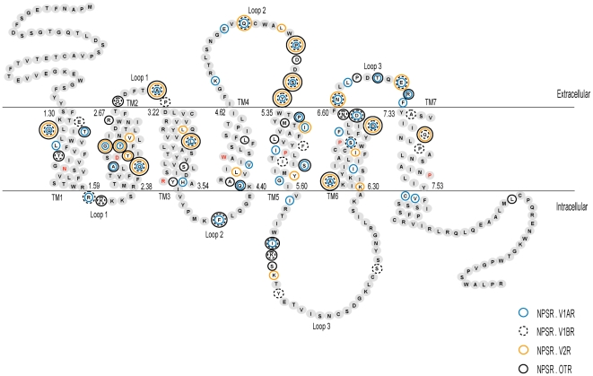
Figure 2. Schematic diagram of the human neuropeptide S receptor.
The sequence is drawn and labeled according to the extracellular, intracellular and transmembrane regions. The boundaries of the three regions were based on the definition of these regions for human NPSR [GenBank: NP_997055] given by the Ballesteros-Weinstein nomenclature and TMHMM program. The most conserved residue in each transmembrane helix is denoted with red text. The first and last amino acid residue numbers in each helix is indicated using Ballesteros-Weinstein numbering scheme. Residues that represent sites of functional divergence between the NPSR and the V1AR, V1BR, V2R and OTR subtypes are marked with outlined circles. Residue-wise functional divergence of NPSR with each subtype of vasopressin-like receptor is provided in Data S3.

