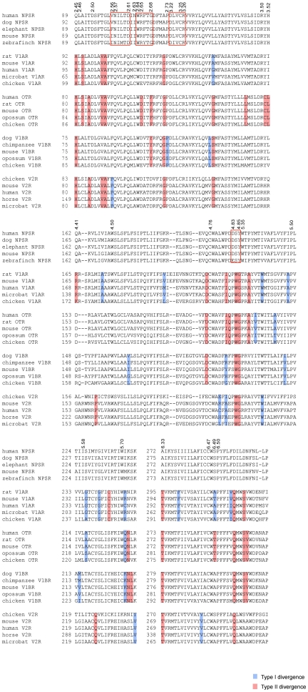
Figure 3. Multiple alignment of functionally divergent sites in the NPSR and vasopressin-like receptors.
Samplings of selected functional divergence-related positions in the region starting from TM2 to the end of ECL3 are shown. Amino acid positions (marked on top) are identified by Ballesteros-Weinstein numbering corresponding to the residue position in human NPSR. Contiguous blocks of conserved residues in the NPSR are shown within hollow boxes. Residues associated with Type I and II divergence are marked in blue background and red background, respectively (Data S3).

