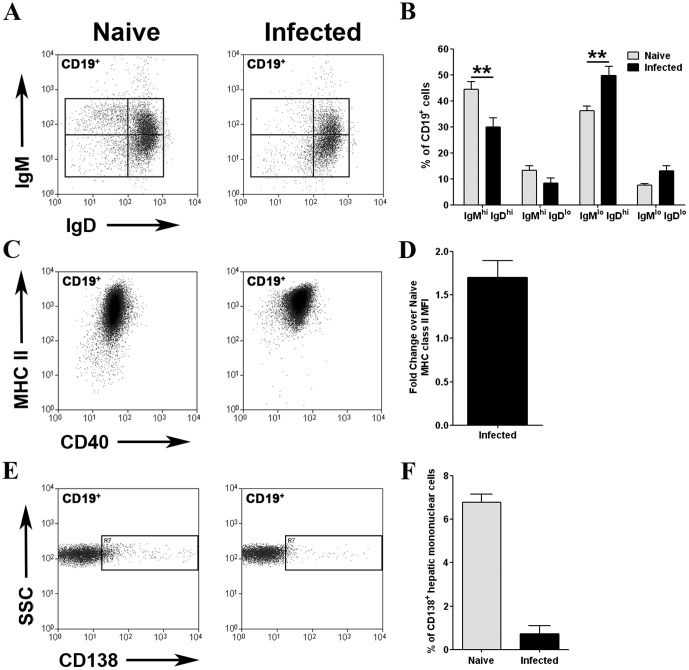
Figure 3. Phenotype of hepatic B cells in L. donovani-infected mice.
A. Hepatic mononuclear cells from naïve and d21 L. donovani-infected C57BL/6 mice were stained for CD19 along with membrane IgM and membrane IgD. B. Frequency of T1, T2, mature and class-switched B cells in naïve (grey bars) and infected (black bars) mice. Data represents the mean ± SEM for 5 independent experiments with cells pooled from 3 mice per experiment (**, p<0.01). C. CD19+ cells were stained for MHCII and CD40. D. Data are shown as fold change in MFI compared to naïve (mean ± SEM from 5 independent experiments). E. Expression of CD138 on CD19+ cells from naïve and infected mice. F. Data are shown as % CD19+ cells in naïve (grey bars) and infected (black bars) mice expressing CD138.

