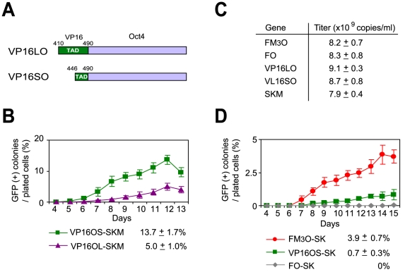
Figure 5. Comparison of the efficiency of making mouse iPSCs with FM3O, VP16SO and VP16LO using Protocol B.
(A) Schematic drawings of the VP16-tagged Oct4s, called VL16LO and VL16SO. Numbers indicate amino acid positions of VP16. (B) Formation of iPSCs from MEFs with two VP16-Oct4 fusion genes and SKM. (C) Virus titer of each construct measured with quantitative RT-PCR. Mean ± SEM from three independent experiments is shown. (D) Formation of iPSCs from MEFs without c-Myc.

