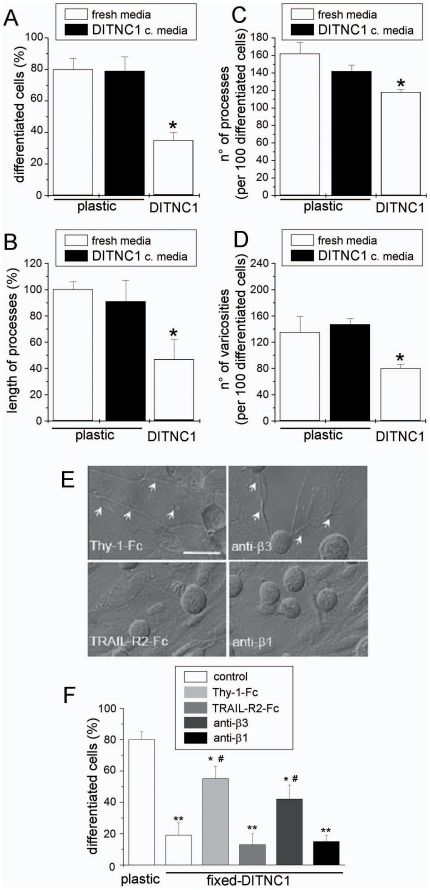
Figure 1. Integrin αVβ3 expressed by DITNC1 astrocytes inhibits neurite extension of CAD cells.
(A–D) Quantification of four different morphological parameters using IMARIS software (Bitplane, Switzerland) of bright-field microscopy images of CAD cells seeded over plastic, seeded over plastic in serum-free medium previously conditioned by DITNC1 cells for 24 hours (black bars) or over a monolayer of DITNC1 astrocytes in serum-free medium. (A) Percentage of differentiated CAD cells with processes ≥15 µm; (B) length of the processes extended by differentiated cells, expressed as a percentage of the control over plastic; (C) number of processes in 100 differentiated cells and (D) number of varicosities per 100 differentiated cells. (E,F) CAD cells seeded over fixed-astrocyte monolayers were induced to differentiate. To block αVβ3 integrin, fixed-cells were incubated with recombinant Thy-1-Fc or antibodies against β3 integrin. TRAIL-R2-Fc or antibodies against β1 integrin were used as controls. Co-cultures were photographed (E) and the percentage of differentiated CAD cells (F) was quantified as in (A). Arrows in E indicate axon-like neurites growing over the DITNC1 mololayer. All graphs show mean+s.e.m. determined from at least 100 cells per condition; n = 3. **P<0.01 or *P<0.05 compared with control cells seeded over plastic. #P<0.05 compared with their respective control.

