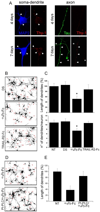
Figure 3. Recombinant αVβ3-Fc inhibits extension of dendrites in cortical neurons.
(A) Cortical neurons, cultured for 4 and 7 days of culture in vitro were stained for Thy-1 (red), MAP-2 (soma/dendrite staining, blue) and Tau (axon staining, green). Arrows indicate some areas with Thy-1 staining. Asterisks label Thy-1-negative axons. (B–E) Neurons, cultured for 4–5 days in vitro were treated with supernatants containing αVβ3-Fc fusion protein (αVβ3-Fc), αVβ3-Fc-depleted supernatants (DS), αVβ3-Fc-depleted supernatants supplemented with TRAIL-R2-Fc (TRAIL-R2-Fc), or non-treated (NT) for 72 hours. Where indicated, 1 U/ml of PI-PLC was added prior to integrin addition (D,E). (B,D) Inverted fields of MAP-2 fluorescence images that were used to count neurons and evaluate dendrite length. (C,E) Quantification of two different morphological parameters performed using Neuro ImageJ software. Results shown are the mean+s.e.m. of 720 neurons from three independent experiments (C) or 240 neurons from two independent experiments (E). *P<0.05 compared with DS condition.

