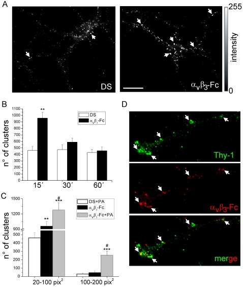
Figure 7. Soluble αVβ3-Fc induces Thy-1 cluster formation on the plasma membrane of living cortical neurons.
Mature cortical neurons were treated with supernatants containing αVβ3-Fc fusion protein (αVβ3-Fc) supplemented or not with Protein A (PA), or with αVβ3-Fc-depleted supernatants (DS) for 15–60 minutes. Neurons were immunostained for Thy-1 only (A) or for Thy-1 and αVβ3-Fc (D). (A and D) Thy-1 clusters (arrows in A) and co-localization with bound αVβ3-Fc (arrows in D) of representative images captured with an epifluorescence microscope are shown. Bar = 20 µm. (B) Data plotted as time versus the number of clusters were obtained from images processed using ImageJ. Ranges of cluster size from 20 to 100 pix2 (1 pix2 = 0.01 µm2). (C) Data plotted as a range of cluster sizes versus the number of clusters for each indicated condition. Results show mean+s.e.m. (12 neurons per condition, n = 6). **P<0.01 and ***P<0.001 compared with their respective control cells in DS at time 15 minutes. #P<0.05 between αVβ3-Fc-Protein A and αVβ3-Fc (D).

