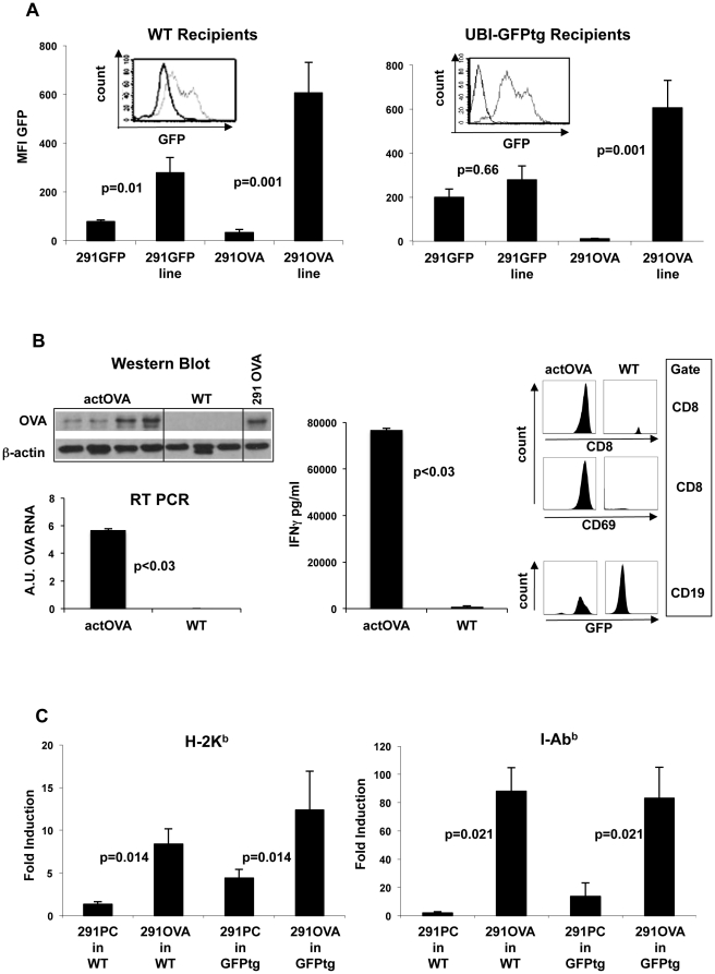
Figure 3. Transfer of OVA-expressing tumor cells into wild-type mice results in loss of antigen and induction of MHC on lymphoma cells.
(A) Systemically growing lymphomas isolated from the spleen after s.c. inoculation of 291GFP or 291OVA cells were analyzed for GFP expression as a marker for antigen expression in comparison to the inoculated cell lines. Introduction of either GFP alone (291GFP) or OVA-IRES-GFP (291OVA) into lymphoma cells resulted in loss of or strong decrease in GFP expression in outgrowing lymphomas after transfer into wild-type mice (left panel, n = 6). Outgrowing lymphomas in GFP-transgenic recipients (tolerant for GFP) likewise lost GFP expression after inoculation of 291OVA cells, but retained the antigen when 291GFP cells had been inoculated (right panel). Flow cytometric histogram inlets show representative examples. Grey lines represent GFP expression at the time of injection, black lines after harvest of lymphomas from spleen. (B) Left: western Blot and RT-PCR analysis (arbitrary units, A.U.) of lymphomas harvested after s.c. inoculation of 291 OVA cells from either OVA tolerant (actOVA) or wild type (WT) recipients. In contrast to WT recipients OVA tolerant actOVA animals did not select for antigen (OVA) loss variants. Middle: Explanted lymphomas were cocultured with unprimed OT-I cells (1∶1 ratio). OVA positive lymphomas from actOVA recipients activated OT-I T-cells to secrete large amounts of IFN-γ (ELISA of supernatant, n = 4, Mann Whitney test) whereas OVA negatively selected lymphomas form wild type recipients did not stimulate OT-I cells. Right: Coculture of OT-I T-cells with lymphoma cells dervived from actOVA or wild type recipients resulted in induction of the T-cell activation marker CD69 expansion of CD8+ (OT-I) cells and reduction of the number of GFP expressing cells CD19+ lymphoma cells. Representative analysis from lymphoma cells harvested from individual mice. (C) Inoculation of 291OVA cells, but not of 291PC cells, into wild-type (n = 5) and GFP-transgenic mice (n = 5) significantly induced MHC class I (left panel) and class II (right panel) in outgrowing lymphoma cells. The strong increase in MHC class I and II expression in outgrowing lymphomas depended on the presence of OVA in the lymphoma inoculum (Mann Whitney test). Fold induction was calculated by comparing MHC expression on freshly explanted lymphoma cells with the corresponding cell line on the same day.

