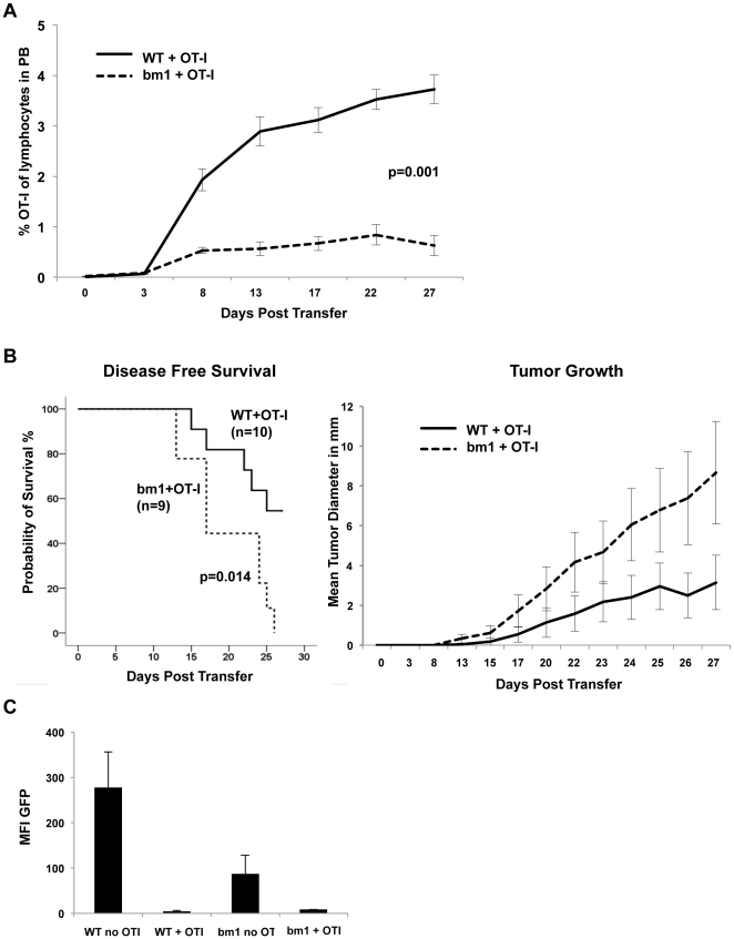
Figure 7. Defective cross-presentation in cells of the recipient impairs T cell response and enhances lymphoma growth.
Recipient animals were T cell depleted as described in Materials and Methods and T cell depletion was continued for 28 days. Wild-type (WT) or bm1 mutant recipient mice received 1×10⁵ 291OVA lymphoma cells s.c. together with 1×10⁶ in vivo primed OT-I cells. (A) Development of an OVA-specific T cell response is impaired in bm1 recipients. Peripheral blood of animals inoculated with 1×10⁵ 291OVA and 1×10⁶ OT-I cells were analyzed by flow cytometry for the presence of CD90.1-positive cells (OT-I) on the days indicated. Numbers of OT-1 T cells are expressed as percentage of lymphocytes in the peripheral blood. In wild-type mice (solid line, n = 10) adoptively transferred OT-I cells expand more readily (Mann Whitney test) than in bm1 recipients (dashed line, n = 9, p = 0.001 for day 8–27). (B) Disease-free survival and cumulative tumor growth after lymphoma transfer: bm1 recipient mice (dashed line) developed tumors significantly faster and cumulative lymphoma growth was enhanced (right panel). (C) Mean fluorescence of GFP in lymphomas arising in wild-type mice or bm1 mice in the presence and absence of OT-I cells. T cell depletion in the absence of OT-I cells (WT no OT-I, n = 9) resulted in preservation of antigen expression, while adoptive transfer of OT-I cells (WT+OT-I, n = 5) led to selection of antigen-negative lymphoma cells.

