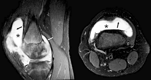
Fig. 2.
Sagittal and axial T2-weighted fat-saturated MR images (TR/TE, 4,000-6,000/30, TR/TE, 2,900-4,300/50 respectively) obtained in a 16-year-old male (group 1, active disease) show synovial hypertrophy (black arrows) and marked effusion (*) in the suprapatellar recesses, and synovial hypertrophy (white arrow) adjacent to the medial posterior condyle

