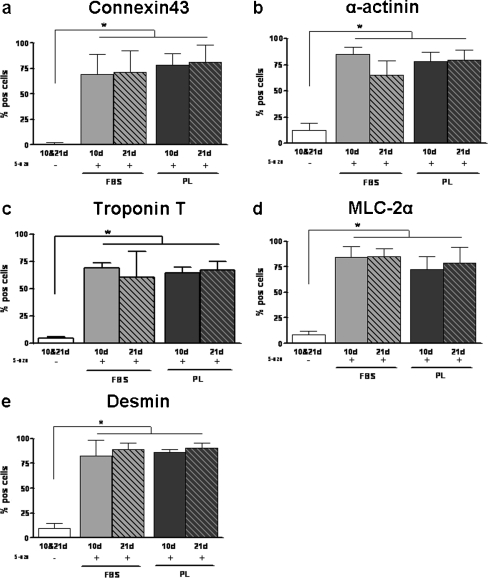
Fig. 6.
Differentation of FBS- and PL-cultured ASC towards cardiomyocytes. Number of FBS- and PL-cultured ASC positive cells for connexin43 (a), α-actinin (b), troponin T (c), MLC-2α (d), and desmin (e) when unstimulated (−) or stimulated with 5-aza-2-deoxycytidin ASC at days 10 and 21. Results are compared with non- stimulated ASC. No significant differences in presence of cardiac proteins were found between FBS- and PL-cultured ASC. Bars means ± SD. n = 6; *p < 0.05

