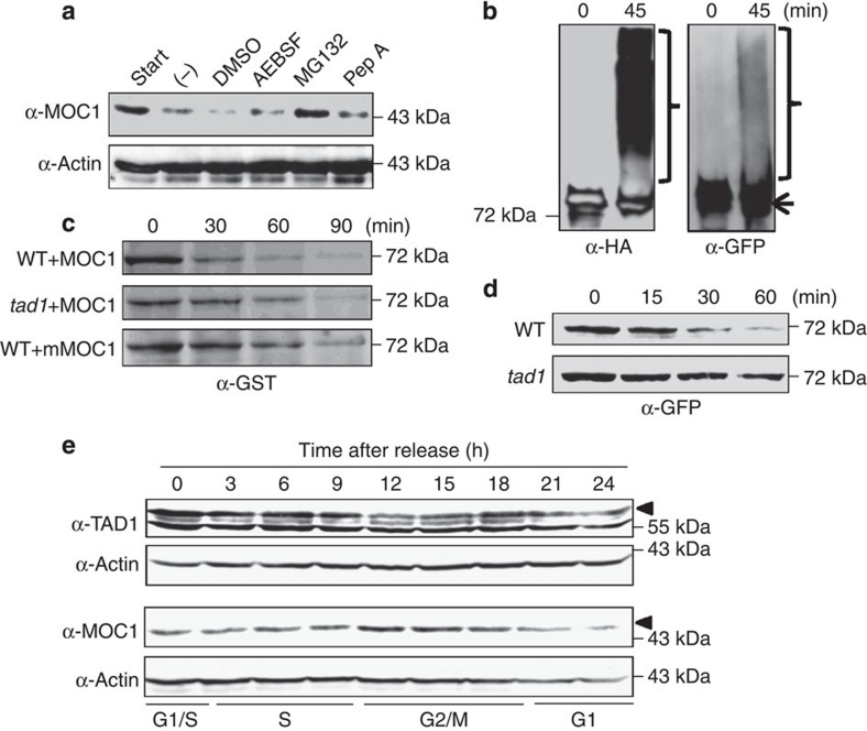
Figure 5. TAD1 targets MOC1 in a cell-cycle phase-dependent manner.
(a) Cell-free degradation assay showing the proteasome-dependent degradation of MOC1. Detection of Actin served as an internal control. (b) In vitro ubiquitination assay of immunopurified MOC1-GFP in the presence of recombinant HA–ubiquitin. The arrow represents unmodified MOC1-GFP, and the brace refers to ubiquitination-modified MOC1-GFP. (c) In vitro cell-free degradation assay showing the delayed degradation of expressed recombinant GST-MOC1 in tad1 and GST-mMOC1 in the wild type. The protein levels at different time points were detected by western blotting using the GST antibodies. (d) Dynamic degradation of MOC1-GFP in the wild-type (WT) and tad1 transgenic calli. Immunoblotting by the GFP antibodies showed the remaining protein levels of MOC1-GFP at different time points. (e) Profiles of TAD1 and MOC1 protein levels during the cell-cycle progression. The cell-cycle phase is determined by flow cytometry at different time points after release from synchronized rice suspension cells as indicated in the scheme at bottom. Black triangles refer to the TAD1 or MOC1 protein in the immunoblots, respectively.

