Figure 1.
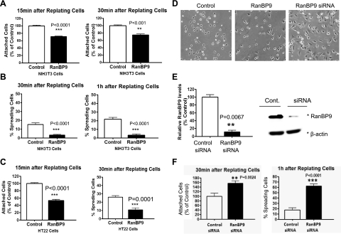
RanBP9 strongly impairs cell adhesion. A, C) NIH3T3 (A) and HT22 (C, left panel) cells were transiently transfected for 24 h, and cells were replated onto fibronectin-coated 96-well plates and incubated at 37°C for 15 or 30 min, after which unattached cells were removed by shaking and tapping, and the amounts of attached cells were quantitated by cresyl violet colorimetric reading. Note that RanBP9 transfection delays cell adhesion to fibronectin-coated plates (A; C, left panel). Values are normalized to vector control-transfected plates. B, C) Quantitation of spreading assays demonstrates markedly reduced cell spreading in RanBP9-transfected NIH3T3 (B) and HT22 (C, right panel) cells; y axis represents the actual percentage of cells spreading after 30 and 60 min. D–F) After RanBP9 siRNA transfection (E), cell attachment and spreading assays were performed (F). Note that knockdown of RanBP9 robustly enhances cell attachment and spreading (F). For spreading assays, images were acquired with a phase-contrast light microscope (Nikon) every 30 min after replating on fibronectin-coated plates (D). Representative images at 1 h after replating are shown. Asterisks at right of immunoblots (E, right panel) indicate protein band positions. Error bars = se. **P < 0.01; ***P < 0.001.

