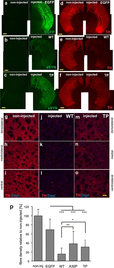
Fig. 6.

Loss of TH-positive innervation in the striatum after expression of a fibrillar αSynuclein variant (WT) or the prefibrillar variant TP. Representative images illustrating the axonal fiber innervation in the striatum 14 weeks after AAV-mediated gene transfer into the SN. Overview of coronal sections showing transgene expression of EGFP (a), WT αSynuclein (b), and TP αSynuclein (c). Nigrostriatal fiber terminals in the striatum were filled with the transgenic protein. Immunohistochemical staining for TH (d–f) revealed that in contrast to the AAV-EGFP-injected animals (d), the expression of αSynuclein led to the appearance of degenerative changes and a loss of striatal fibers, which was more pronounced for WT (e). Scale bar 500 μm. g–o Higher magnification images covering the dorsal, medial and ventral area of a representative striatal section of an intact non-transduced control side (g–i), WT-transduced (j–l), and TP-transduced (m–o) brain. TH-immunostained sections showing the extent of loss of striatal innervation. Remaining fibers in the WT αSynuclein-treated animals were sparse. Scale bar 100 μm. p Quantification of fiber density revealed a clear reduction in TH-positive striatal innervation in the αSynuclein transduced animals at 14 weeks after injection (significant difference from the EGFP-transduced control group at p < 0.001). The difference between the WT and TP variant was also significant (p = 0.048), whereas the difference between A30P and TP was not
