
Figure 3. Effect of platelet PN-1 in PRC lysis.
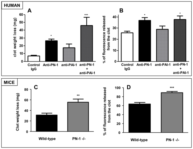
(A–B) PRCs from PRP of healthy donors, in the presence of an irrelevant IgG or an anti-PN1 IgG, or an anti-PAI-1 IgG or (C–D) PRCs from WT or PN-1−/− mice PRP, were incubated with FITC-fibrinogen prior to clot formation. (A, C) The percentage reduction in clot weight and (B, D) the percentage of released fluorescence were analyzed over 24 hours. Data are presented as means ± SEM of 5 independent experiments from different donors and mice. ***P < 0.001, significantly different from control IgG or WT clots. *P < 0.05, significantly different from control IgG. **P < 0.01, significantly different from WT clots.