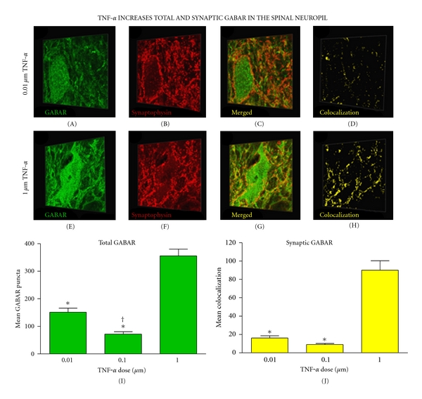
Figure 4.

Increased synaptic GABAAR expression in the neuropil 60 min after TNFα injection. (a)–(h) Three-dimensional representative images of motor neurons demonstrating a dose-dependent increase in synaptic GABAARs in the neuropil after TNFα injection. (i) Quantification of confocal stacks shows a significant increase in total GABAARs following the highest dose of TNFα (*P < 0.001 from middle and lowest doses). An increase in total GABAARs was also observed in the lowest dose relative to the middle dose († P = 0.015). (j) An increase in synaptic GABAARs occurred following the highest dose of TNFα (*P < 0.001 from middle and lowest doses). Bars represent group means across >800 confocal image stacks (12 subjects, n = 4 subjects per group). Error bars reflect SEM. Scale bar, 30 μm.
