
Figure 1.
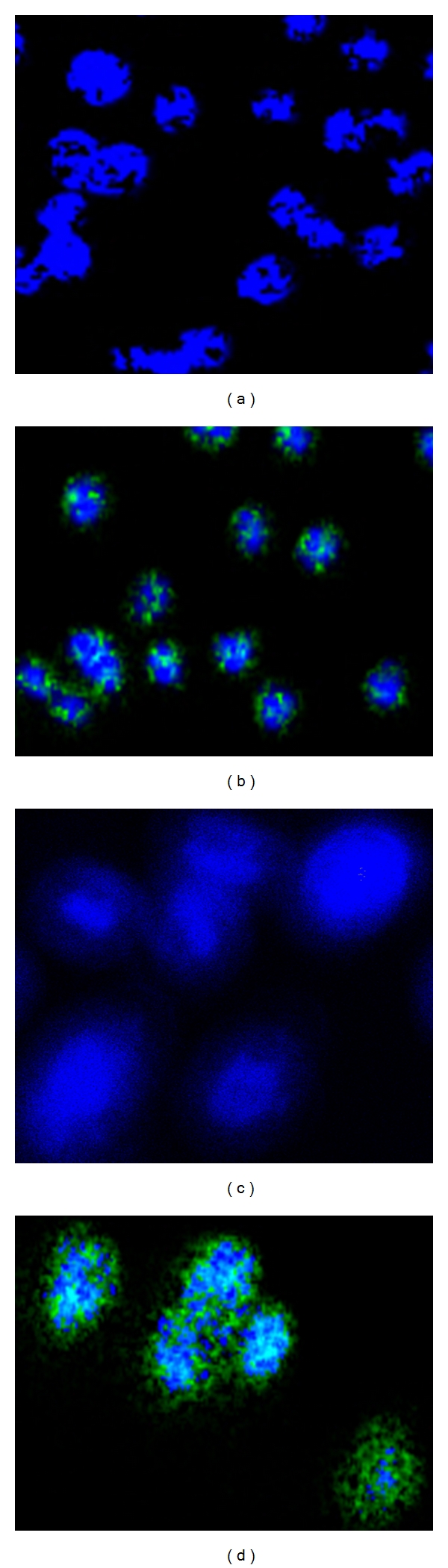
Photomicrographs showing infectivity of cells with C. trachomatis . Mouse J774 macrophages (a and b) and HeLa (c and d) (2.5 × 10⁴ cells/well) were infected with C. trachomatis EBs (2.5 × 10³ IFU/well) and incubated for 2 days at 37°C and 5% CO2. Harvested cells were stained with a monoclonal antibody to C. trachomatis and an FITC-labeled secondary antibody (green) and the nuclei counterstained with DAPI (blue). Chlamydia was visualized by confocal fluorescence microscopy.