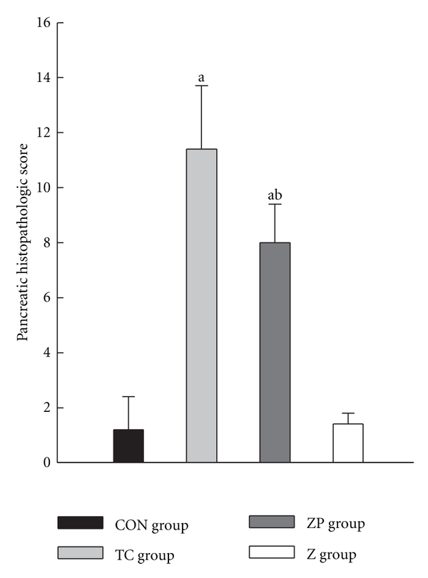
Figure 4.

Pancreatic histopathological scores in all groups. The pancreatic tissue was with hematoxylin-eosin staining. The sections were evaluated by two independent pathologists who were blinded to this research. Time course changes of the pancreatic histological assessment were scored based on edema, inflammation, hemorrhage, and necrosis according to the scale described by Schmidt et al. [19]. a P < 0.05 versus CON group. b P < 0.05 versus TC group.
