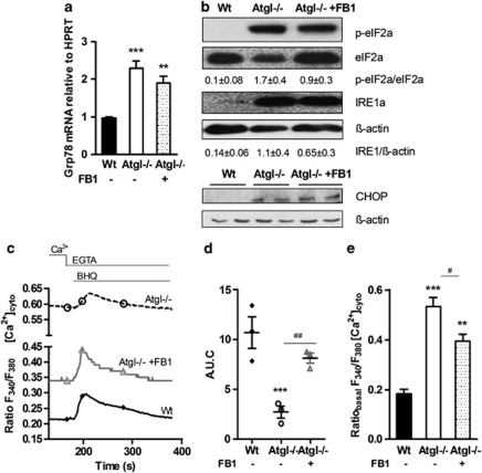
Figure 6.
Persistent ER stress after inhibiting ceramide synthesis in Atgl–/– macrophages. (a) mRNA expression of Grp78/BiP in Wt, Atgl–/– and FB1-treated Atgl–/– macrophages, including normalization to hypoxanthine-guanine phosphoribosyltransferase (HPRT), was determined by real-time PCR. **P≤0.01, ***P≤0.001. (b) Cytosolic fractions of macrophages were isolated and proteins were resolved by SDS-PAGE. Protein expression was determined using specific antibodies for phosphorylated (p)eIF2α, eIF2α, and IRE1α by western blotting. Data are expressed as the ratios of eIF2α/eIF2α and IRE1α/β-actin of two independent experiments±S.E.M. CHOP protein expression was analyzed in whole cell lysates. The expression of β-actin was determined as loading control. (c) Wt, Atgl–/– and FB1-treated Atgl–/– macrophages were plated on coverslips in DMEM/10% LPDS. The fura-2 fluorescence ratio (340/380 nm) was determined in single macrophages before and after the addition of BHQ (15 mM) in the presence of 1 mM EGTA. Data are presented as mean values±S.E.M. of 300 cells per genotype of three independent experiments. (d) Area under the curve after the addition of BHQ was calculated. Dots represent means of 300 cells of three independent experiments±S.E.M. ***P≤0.001; ##P≤0.01. (e) Basal cytosolic Ca2+ concentrations were calculated from the fura-2 fluorescence ratios (340/380 nm) during the initial 5 min. **P≤0.01, ***P≤0.001; #P<0.05

