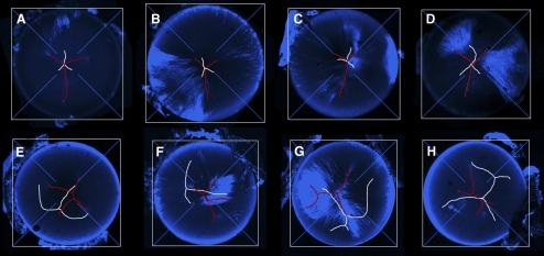
Figure 6.
Gallery of images showing lens suture organization in 8-week-old wild-type (A–D) and Epha2−/− (E–H) mice. Anterior sutures are shown in red and posterior sutures in white. The optical axis is located at the convergence of the crosshairs. Images are maximum intensity orthographic projections of FM 4-64–stained lenses. In wild-type lenses, the sutures are located on the optic axis and have a characteristic Y-configuration. In contrast, sutures in Epha2−/− animals are not centered on the optic axis and exhibit a range of aberrant morphologies.

